Understanding and re-designing cell division machinery
We study the machines and mechanisms that help eukaryotic cells achieve accurate genome inheritance. Using these insights alongside de novo protein design, we reimagine key mechanisms and reverse engineer new machines for synthetic biology. Our research stands at the interface of cell biology, synthetic biology, biophysics, and biochemistry — from single molecules to living cells.
Three interconnected frontiers
AI-aided de novo protein design for engineering new cytoskeletal proteins
We harness generative AI tools — RFdiffusion, ProteinMPNN, AlphaFold — to design novel cytoskeletal proteins from scratch. Our goal is to build synthetic cytoskeletal systems with tunable mechanical and dynamic properties.
Read morePerturbation and adaptation of the mitotic checkpoint in cancer biology
We build detailed mathematical models of the mitotic checkpoint's signaling cascade using quantitative in vivo measurements — relying on CRISPR-engineered cell lines and live-cell fluorescence microscopy.
Read moreReverse engineering kinetochores using de novo-designed proteins
Building on decades of kinetochore research, we pioneer efforts to reverse engineer this machine using de novo-designed proteins — constructing simplified kinetochore-like machines for synthetic biology.
Read moreWhat's happening
Welcome, Riley Zheng and Mia Levin!
We are excited to welcome Riley Zheng and Mia Levin to the lab as undergraduate research assistants. We look forward to working with you!
Anish Virdi recognized as Outstanding Undergraduate Student
Anish Virdi has been recognized as the Outstanding Undergraduate Student by the Biophysics class of 2026. Congratulations, Anish, on this well-deserved honor!
Soubhagyalaxmi Jema receives Barbour Scholarship
Soubhagyalaxmi has been awarded the U-M Rackham Graduate School Barbour Scholarship, which supports academic excellence among women from Asia and the Middle East pursuing advanced degrees at U-M.
Jema's paper published in Current Biology
A critical new finding on spindle assembly checkpoint signaling! Congratulations Soubhagyalaxmi on this important contribution.
View on PubMedRoy et al. paper published in eLife
New work on MELT motifs in Spc105 that balance the strength and responsiveness of the spindle assembly checkpoint.
View on PubMedUS patent issued for eSAC technology
U.S. Patent No. 15/355,824 — "Activating Mitotic Checkpoint Control Mechanisms." Describes the eSAC method for controlling the duration of cell division using designed protein fragments.
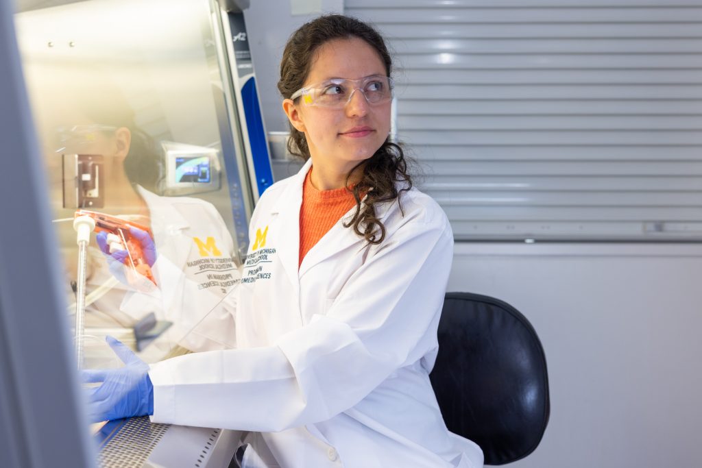
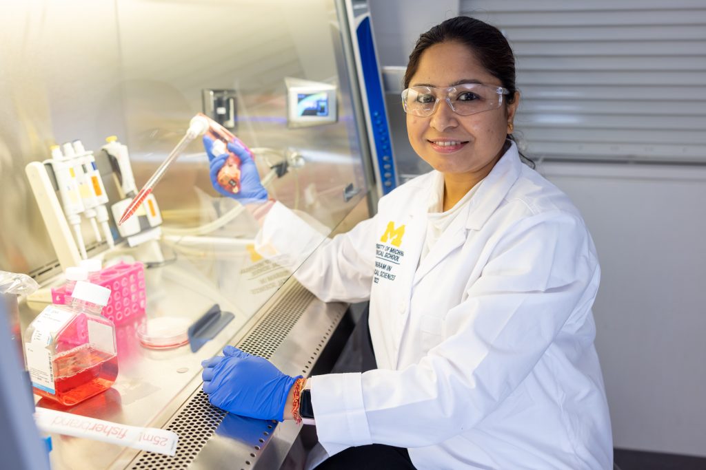
Lab members
We are a collaborative, interdisciplinary team of scientists passionate about understanding cell division.

Ajit Joglekar
Professor · PI
Lina Pena
Graduate Student · Biophysics

Yiwei (Ryann) Li
Graduate Student · CDB
Soubhagyalaxmi Jema
Graduate Student · CDB
Anish Virdi
Undergraduate Student
Jennifer Guan
Undergraduate Student
Sydney Lee
Undergraduate Student
Riley Zheng
Undergraduate Student
Mia Levin
Undergraduate Student
Dubuke Ma
Technician
How to Train Custom Cell Segmentation Models Using Cell-APP. ↗
Training accurate cell segmentation models typically requires large annotated datasets and deep computational expertise—resources inaccessible to most cell biology labs. This protocol provides step-by-step instructions for using Cell-APP to train custom deep-learning segmentation models from user-generated microscopy images, requiring no programming background. The workflow covers image acquisition guidelines, annotation strategies, model training, and validation, with troubleshooting guidance for common failure modes. The protocol is designed to make state-of-the-art cell segmentation broadly accessible to biologists working with diverse cell types and imaging modalities.
PMID 41769259Cell-APP: A generalizable method for cell annotation and cell-segmentation model training. ↗
Accurate segmentation of cells in microscopy images is a fundamental bottleneck in quantitative cell biology, yet building custom deep-learning models typically demands significant computational resources and expertise. Cell-APP is a generalizable pipeline that combines automated segmentation with a user-friendly annotation interface, enabling researchers to generate training datasets and fine-tune models for their specific cell type and imaging conditions. The method was validated across multiple imaging modalities and cell types, substantially reducing the manual effort required for large-scale microscopy analysis. Applied to chromosome segregation imaging, Cell-APP enables systematic, high-throughput analysis of kinetochore and spindle dynamics.
PMID 39896521The structural flexibility of MAD1 facilitates the assembly of the Mitotic Checkpoint Complex. ↗
The spindle assembly checkpoint (SAC) relies on rapid assembly of the Mitotic Checkpoint Complex (MCC) at unattached kinetochores to halt cell division until all chromosomes are correctly bi-oriented. This study reveals that the middle domain of MAD1—long thought to be a simple coiled-coil spacer—undergoes significant conformational flexibility that is critical for MCC formation. Using FRET measurements and biochemical reconstitution, the authors show that MAD1 flexibility allows it to simultaneously engage both MAD2 and the BUBR1–BUB3 heterodimer in a multivalent fashion. This flexible architecture explains how a single MAD1 dimer can efficiently nucleate MCC assembly even when kinetochore-bound MAD1 levels are limiting.
PMID 36934097Signaling protein abundance modulates the strength of the spindle assembly checkpoint. ↗
The spindle assembly checkpoint must be calibrated to robustly delay mitosis when chromosomes are unattached, yet allow timely progression once all kinetochores achieve correct attachments. This study demonstrates that the absolute abundance of SAC proteins—particularly Bub1 and BubR1—modulates checkpoint strength in a graded manner across individual cells. By quantifying protein levels and correlating them with checkpoint duration, the authors show that natural cell-to-cell variation in protein copy number produces corresponding variation in checkpoint robustness. A mathematical model further shows that protein abundance tunes the system near a threshold, making the SAC sensitive to stoichiometric changes rather than simply switching between on and off states.
PMID 37738972Aurora B phosphorylates Bub1 to promote spindle assembly checkpoint signaling. ↗
Bub1 kinase at kinetochores is essential for generating the SAC wait-anaphase signal, but how its activity is regulated in response to attachment status was unclear. This study shows that Aurora B—the error-correction kinase of the chromosome passenger complex—directly phosphorylates Bub1 at kinetochores to enhance SAC signaling. Phosphorylated Bub1 exhibits stronger interaction with Mad1, thereby promoting MCC assembly at unattached and incorrectly attached kinetochores. These findings reveal how the cell mechanistically links two major kinetochore activities—error correction and checkpoint signal generation—through a shared upstream kinase.
PMID 34861183Kre28–Spc105 interaction is essential for Spc105 loading at the kinetochore. ↗
Spc105 (KNL1 in humans) is the key outer kinetochore scaffold that recruits checkpoint proteins via its MELT repeats, yet how Spc105 itself is loaded onto the kinetochore has been poorly understood. This study identifies a direct interaction between Kre28—a subunit of the budding yeast DASH/Dam1 microtubule-binding complex—and the N-terminal region of Spc105. Disrupting this interaction causes a severe reduction in Spc105 levels at kinetochores and compromised checkpoint signaling. These findings reveal a new pathway for outer kinetochore assembly in which the microtubule-binding apparatus directly recruits the checkpoint scaffold, functionally coupling spindle attachment with checkpoint competence.
PMID 35042402BubR1 recruitment to the kinetochore via Bub1 enhances spindle assembly checkpoint signaling. ↗
BubR1/MAD3 contributes to MCC formation and can be recruited to kinetochores through multiple mechanisms. This study demonstrates that indirect BubR1 recruitment via Bub1 is a more potent enhancer of SAC signaling than direct kinetochore recruitment through other interactions. Combining quantitative live-cell imaging with mathematical modeling, the authors show that Bub1-dependent BubR1 recruitment amplifies local MCC production, enabling robust checkpoint signaling even when kinetochore occupancy is limiting. This indirect recruitment strategy—using a scaffold to concentrate a downstream effector—may represent a general principle for amplifying signaling at macromolecular assemblies.
PMID 35767360The human kinetochore links chromosomes to spindle microtubules through a complex assembly of more than 100 proteins, yet its precise architecture under physiological conditions was largely unknown. This study uses intramolecular FRET measurements with site-specifically labeled proteins to map the positions of key kinetochore components in live human cells under different states of microtubule attachment and centromeric tension. The results reveal that microtubule attachment and tension collectively reshape the kinetochore's protein architecture, compacting it and repositioning checkpoint proteins away from their activating kinase. This provides a direct molecular explanation for how successful chromosome bi-orientation mechanically transforms the kinetochore to silence the SAC and permit mitotic exit.
PMID 33035484Spc105/KNL1 contains multiple MELT motifs that are phosphorylated by Mps1 to recruit Bub1 and BubR1 for SAC signaling, yet the copy number and sequence variation among these motifs varies widely across species. This study systematically varies both the number and individual strengths of MELT motifs in budding yeast Spc105 to show that both parameters jointly determine SAC strength and responsiveness to inhibition. High copy numbers of weak motifs produce a checkpoint sensitive to partial disruption, while low copies of strong motifs create a less tunable response. The data suggest that the natural MELT repertoire of Spc105 is optimized to balance robust checkpoint activation with the ability to rapidly silence signaling upon chromosome attachment.
PMID 32479259Understanding the internal logic of the SAC requires tools that activate the pathway outside its normal kinetochore context. This study constructs synthetic "ectopic SAC activation" platforms by tethering individual SAC components to defined cytoplasmic locations, probing the biochemical requirements for MCC formation step by step. By systematically reconstituting signaling in ectopic contexts, the authors delineate which protein–protein interactions are necessary and sufficient for MCC production, independent of kinetochore geometry. The results reveal that the SAC cascade operates as a series of modular steps with distinct requirements, and that catalytic amplification by kinetochore-bound MAD1 is the key driver of rapid MCC production in living cells.
PMID 30595520Protein phosphatase 1 (PP1) bound to Spc105/KNL1 silences the SAC after chromosomes attach to spindle microtubules, but the same Spc105 region also regulates kinetochore–microtubule attachment stability. This study uses quantitative genetics and live-cell imaging in budding yeast to dissect these two PP1-dependent functions. The results show that Spc105-bound PP1 makes distinct, separable contributions to checkpoint silencing and to the regulation of kinetochore–microtubule attachments—mutations that selectively impair one function leave the other largely intact. These findings clarify how a single phosphatase-binding site on the outer kinetochore simultaneously orchestrates two critical determinants of faithful chromosome segregation.
PMID 31649151The kinetochore encodes a mechanical switch to disrupt spindle assembly checkpoint signalling. ↗
How the kinetochore transitions from generating a "wait-anaphase" signal to silencing the checkpoint upon correct microtubule attachment has been a central question in chromosome segregation biology. This study uses FRET-based measurements in living yeast to show that tension generated by microtubule attachment physically stretches the kinetochore, repositioning the checkpoint activator Bub1/Mad1 away from the activating kinase Mps1. The resulting spatial separation constitutes a mechanical switch that autonomously shuts off checkpoint signaling in response to the physical state of chromosome–spindle attachment. This mechanism directly couples kinetochore mechanics to biochemical signaling, revealing how correct attachment is sensed at the molecular level.
PMID 26053220Molecular architecture of a kinetochore-microtubule attachment site. ↗
The kinetochore is the multiprotein complex that attaches chromosomes to spindle microtubules to drive their segregation, but its molecular organization was entirely unknown at the time of this work. Using quantitative fluorescence microscopy in living budding yeast, this study measured the stoichiometries and positions of key kinetochore proteins relative to each other and to the microtubule plus-end. The resulting molecular map revealed an end-on attachment geometry with distinct inner and outer kinetochore layers arranged along the microtubule axis. This study was among the first to define the nanoscale molecular architecture of a kinetochore–microtubule attachment site and established a structural framework that has guided the field for nearly two decades.
PMID 16715078For the complete list, visit the Publications page.